A 60s year old male was doing bending exercises in physical therapy when he had an episode where he felt lightheaded, chest pressure and shortness of breath. He presented to the emergency department as a ‘Code Sepsis.’ Vital signs: BP 101/69, HR 118, afebrile, RR 24, 85% (improved to mid 90s with supplemental 02). In the Resuscitation bay he really wasn’t complaining of infectious symptoms, and was alert and oriented and answering questions appropriately. His right lower extremity was larger than his left. Electrocardiogram and point of care ultrasound images are below.

ECG: no STEMI, sinus tachycardia 118, normal axis, right bundle branch block, ST depression most pronounced V3-4
POCUS Rapid Ultrasound for Shock and Hypotension (RUSH exam) showed a plethoric inferior vena cava (clip 1), a clot in transit in the right atrium (clip 2), and a right lower extremity deep vein thrombosis (clip 3). He was treated immediately for his acute pulmonary embolism and discharged from the hospital a couple weeks later.

Clip 1: Plethoric Inferior Vena Cava with minimal respiratory variation

Clip 2: Right atrial clot in transit seen on subxiphoid window
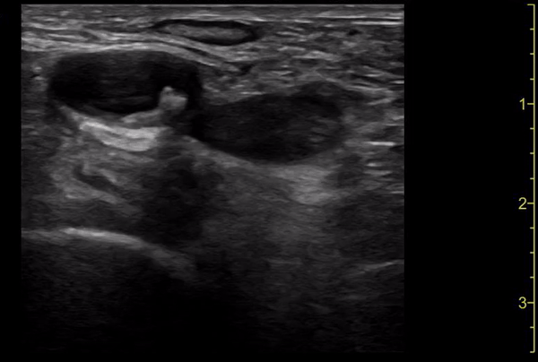
Clip 3: Deep vein thrombosis seen in right lower extremity
Undifferentiated hypotension: Rose, Bair, Mandavia, & Kinser in California described their approach in 2001. Weeks, Zapata, & Napolitano in the Bronx, NYC published the role of sonography in the undifferentiated hypotensive patient in 2007. Weingart, Duque, and Nelson also in NYC in 2009 further described the HI-MAPP approach.
HI-MAPP
Heart
IVC
Morisson’s (which we know now should be caudal tip of liver
Aorta
Pneumothorax/Pipes (DVT)