A 50 something year old female with a past medical history of breast cancer presented for epigastric pain with 2 episodes of pre-syncope over the past couple days. She denied fever/chills, cough, dyspnea, chest pain, GU, GI, or GYN symptoms. Vitals were notable for tachycardia to 119 and hypotension (BP 105/81). Her FSG read as “HI” on initial triage and she was initiated on IV fluids for treatment of suspected hyperglycemia triggered by an infectious source. Initial blood work was notable for ALT 249, AST 150, Alk Phos 131. Given the epigastric pain and transaminitis, a POCUS biliary study was done to see if the gallbladder was the culprit. Exam of the epigastric region, however, quickly revealed pathology outside of the biliary tract.
Anechoic fluid near the heart was seen from the epigastric windows. Providers quickly switched gears, and a POCUS cardiac study revealed a pericardial effusion with a right ventricular diastolic collapse (clips 1 and 2 below; specific for echocardiographic tamponade) and a plethoric IVC (clip 3 below; sensitive for echocardiographic tamponade. [https://doi.org/10.1016/j.ajem.2018.11.004]
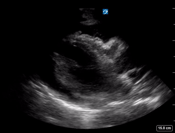
Clip 1: POCUS cardiac parasternal long axis with RV diastolic collapse, the most specific finding for echocardiographic tamponade.
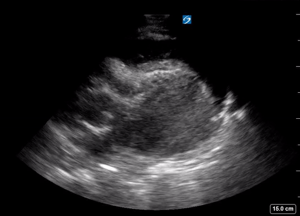
Clip 2: POCUS cardiac subxiphoid window with RV diastolic collapse, the most specific finding for echocardiographic tamponade.
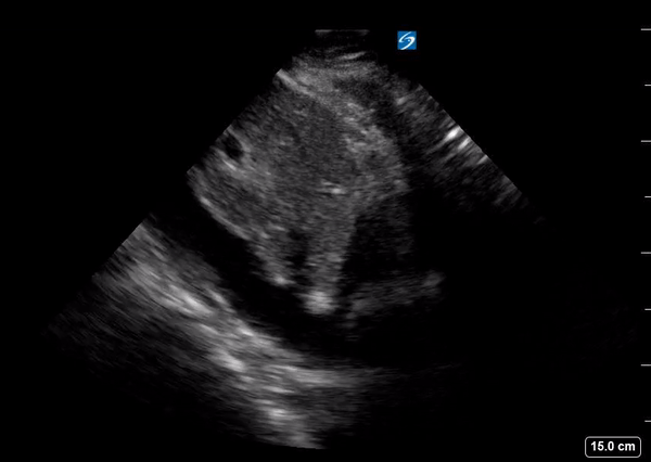
Clip 3: Plethoric IVC with minimal respiratory variation, the most sensitive finding for echocardiographic tamponade.