
Image 1: POCUS thoracic left anterior chest - lung point, highly specific for pneumothorax
A young patient suffers a gunshot wound to the left lateral chest. On arrival, resuscitation ensues, large bore IV access, vital signs, primary survey etcetera.
Performed simultaneously with initial resuscitation, the thoracic views of the EFAST are obtained with a linear transducer (highest resolution). The left anterior chest shows a lung point, the border of the pneumothorax where part of the pleura is sliding, and part is not, a highly specific finding (Image 1 above). The right chest shows good lung sliding, no pneumothorax in that lung field (image 2 below). A portable chest radiograph was significant for a mediastinal location of the bullet, left sided subcutaneous emphysema, but no definitive pneumothorax (image 3 below). A left sided thoracostomy tube was placed with the resolution of the hemo-pneumothorax over several days. The tube was subsequently removed and the patient was discharged in good health.
Pearl: When evaluating the pleural line, find the ribs first, the pleural line lies just deep to the ribs.
Image 2: POCUS thoracic right anterior chest - good lung sliding
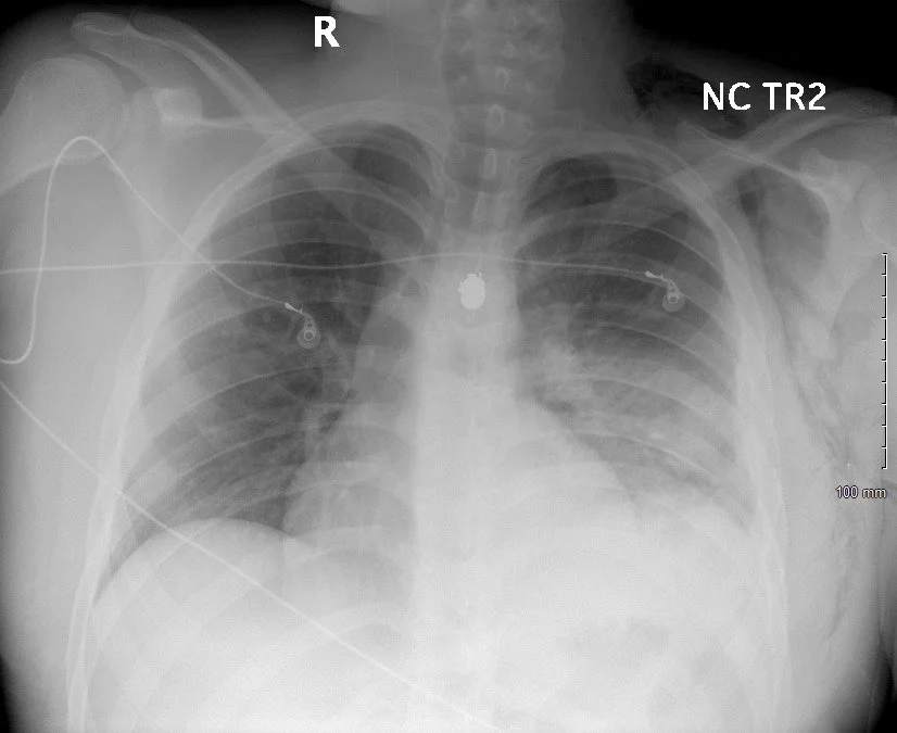
Portable chest radiograph: No definitive pneumothorax, left lateral subcutaneous emphysema, midline bullet fragment