
Clip 1: Apical 4 Chamber view with clot in right atrium moving too-and-fro through the tricuspid valve
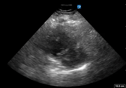
Clip 2: Parasternal Short Axis view showing flattening of the intraventricular septum (D sign) as a result of increased right heart pressures.
A 40s year old female presented to the emergency department with progressively worsening shortness of breath for the past week. No other complaints, says dyspnea getting worse, and now she can’t keep up with her young children. Vital signs were; HR 114 beats/min, BP 134/75 mm Hg, RR 16 breaths/min, Temp 98.6F, 02 saturation 100% on room air.
Cardiac POCUS performed during initial interview revealed a clot in transit (Clip 1), signs of right ventricular strain including D-sign and RV dilation (Clip 2). A heparin infusion was started and within an hour of arrival the patient was transferred to the Cardiac Intensive Care Unit for thrombectomy.
POCUS Pearl: A case to manifest the power of the focused echo in undifferentiated dyspnea.