
Image 1: Aorta above is misidentified as a plethoric IVC

Image 2: True image of the Inferior Vena Cava (IVC)
A very common false positive is the aorta misidentified as a plethoric IVC. The patient in question is short of breath and has a poor EF on POCUS cardiac, and the provider nicely goes to check on the IVC, but images of the aorta are mistakenly obtained (Image 1).
Image 1: The Aorta with its characteristic thick hyperechoic (white) walls.
Image 2: True image of an IVC. Thin walls and traces into the right atrium.
POCUS Pearl: Anatomically, the aorta and IVC run right next to each other, so an easy and common ultrasonographic error to commit. Keep an eye out, practice toggling between the two to help correctly identify and avoid this trap.
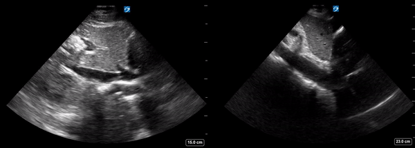
Image 3: Side by side mosaic of IVC (on the Left) and aorta (on the Right)